DICOM Reader – medical imaging software
The software allows you to view the content of the PACS server and external sources.
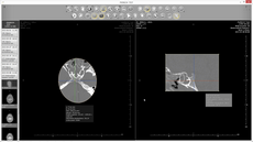
Our DICOM image viewer allows you to display images in diagnostic quality for radiologists as well as reference for clinicians. These images are generated on the Image Distribution servers, which the client is connected to, while the image distribution server enable viewing of the tests from the list of connected PACS servers.
 VIEW APPLICATION CLIP
The architecture of the Image Distribution system has been designed to maximize the use of hardware resources and to reduce the waiting time for the use of the tool, therefore calculations are performed on a dedicated server, which in a minimum configuration is much stronger than the basic workstation located in the branch. This solution allows you to perform advanced 3D reconstructions: MPR, VOL, MIP, minIP, etc. in a much shorter time than it would be on the aforementioned workstation.
Radiology DICOM Viewer Offer
We give you the application for viewing and medical diagnostics of medical tests (CT, MR, X-ray, Mammography, etc.). Exhibeon is a solution addressed to doctors, technicians and is used to search, view and analyze digital photos in the DICOM standard. It works with medical devices supporting DICOM 3.0 communication protocols. Depending on the configuration, the application can operate in two modes: diagnostic and clinical ones. The diagnostic client is intended for radiologists to view the tests at a descriptive station which the corresponding diagnostic monitors are connected to. The clinical client is intended for departmental physicians so that they can review the study at a reference station located, for example, in a ward.
DO system architecture allows for easy and definitely faster description of teleradiological tests. As the application can be made available to physicians directly from hospital servers, there is no need to send a large number of DICOM files to descriptive stations located outside the hospital. As a result, the doctor can describe the study as soon as it is sent from the device to the PACS server.
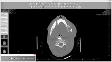
The browser supports medical images in the DICOM 3.0 format. The software can read over 30 varieties of DICOM format, including films – Cine mode.
Application’s features
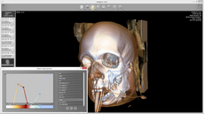
3D
Blood vessel analysis
CINE animation
Automatic virtual colonoscopy
Blenda – Electronic iris

CPR

DSA

Findings

Fusion and image recording

Histogram

HP configurator

Image importing

Magnifying glass

Mammography

ADC maps

Perfusion maps

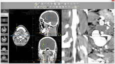
MPR

Numbering ribs

Opening an encapsulated PDF

Perfusion, removing movement artifacts

Measurements

Magnification

Transformations

Layer thickness regulation

Segmentation of the liver

Segmentation of selected organs

User’s session

Spectroscopy

MR image subtraction

Removing table

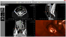
Virtual colonoscopy

Window & Level

Time intensity graphs

Pathological changes – RECIST